Intracellular development of filariae in the vector
During its bloodmeal, the Simulium fly ingests the microfilariae from the skin of their host. At the same time, the infective larvae (L3), if present, escape.
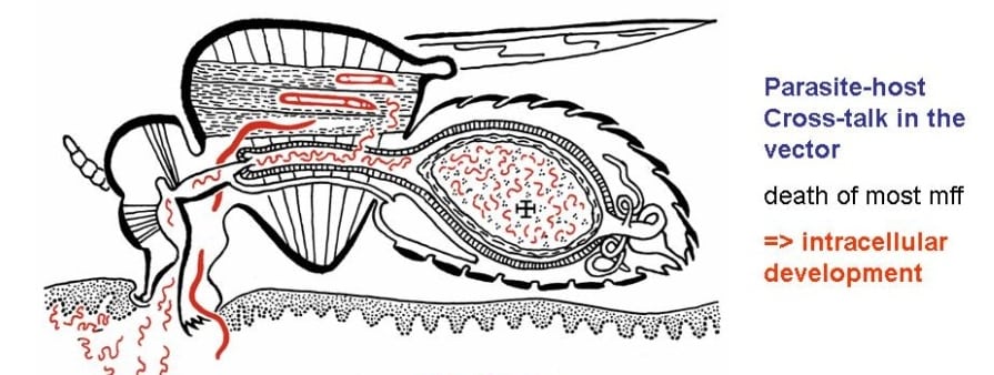
Development of Onchocerca microfilariae in the Simulium vector fly (from Wenk & Renz, Parasitologie)
The development from the microfilaria to the metacyclic infective larva (L3) takes about 7 to 10 days, depending on the ambient temperature.
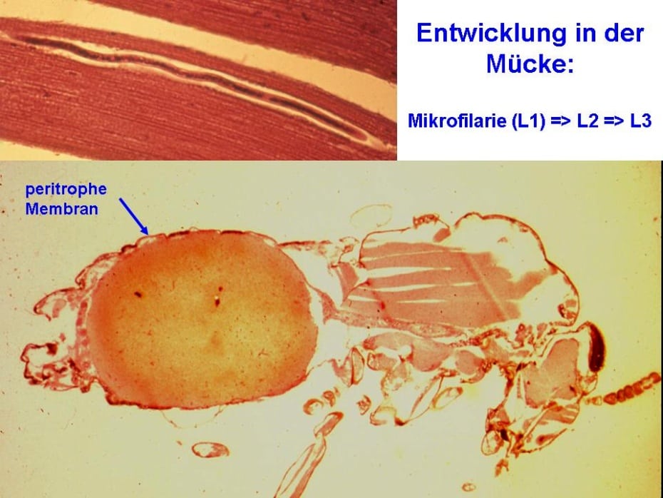
Histological section through a freshly engorged fly (photo Dr. Mohamed Omar, Hamburg)
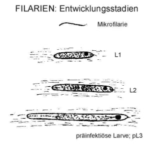
The blood in enclosed by the peritrophic membrane, which is secreted by the cells of the mid gut. This membrane must be penetrated by the microfilaria, before it migrates through the hemolymph and settles in the syncitial cells of the longitudinal flight muscles in the fly thorax. There they grow and moult twice before they reach the infective stage (L3). Development of all filariae, as far studied, is always intracellular and involves the hypertrophy of the parasitized cell.
The second stage larvae is immobile. Its looks as a sausage, hence they are called: ‘sausage-stage larva’.
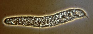
sausage-stage in Simulium damnosum
This sausage-stage larva, however, is dead and disintegrating, as one can see from the damaged surface of its cuticula.
Onchocerca larvae in the flight muscle (A. Renz)
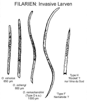
Infective larvae found in Simulium damnosum in Cameroon (A. Renz)
Since many years, different ‘types’ of infective larvae were found and distinguished in wild-caught Simulium flies, dissected in Norther Cameroon (Duke, 1967, Renz 1987). They were named: Type A, B, etc. Presumably not all are true Onchocerca species (Type F, and H!). Type G was identified being Onchocerca ochengi (Wahl, Ekale, Enyong, Renz, 1991).
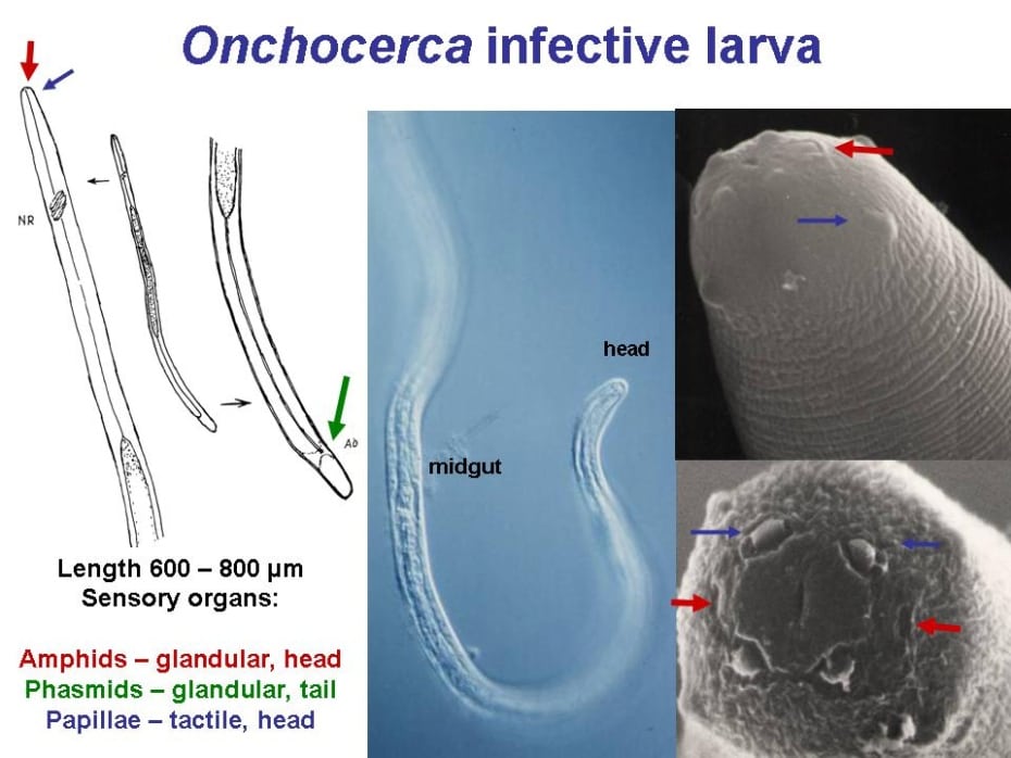
Infective L3 larvae of Onchocerca. (REM-photo of L3-heads from Franz & Renz, 1980)
Although the infective larvae of many Onchocerca species look rather similar – and have been mixed up frequently in the past! – they can be distinguished morphologically by their length, the shape of their anterior end and tail. To be sure, we now use molecular techniques to amplify their DNA and sequence the PCR-product.


Leave a Reply
Want to join the discussion?Feel free to contribute!